Liposomal NMN: Liposome Technology, NAD+ Response and Formulation Insights
What Is a Liposome?
Liposomes were first described by Bangham and colleagues in 1965. They are microscopic vesicles formed by lipid bilayers and have a membrane-like architecture: hydrophilic regions face the aqueous environment, while hydrophobic regions are located within the bilayer.
This amphiphilic structure allows liposomes to accommodate water-soluble compounds in aqueous regions and lipid-soluble compounds within the lipid bilayer. For nutrient delivery, the liposome can therefore act as a protective carrier around an active ingredient.
How a Liposome Forms
Phospholipids are amphiphilic molecules with a hydrophilic head and hydrophobic tail. When dispersed in water, they can self-assemble into bilayer structures as hydrophobic regions orient away from the aqueous phase. The bilayer can then close into a vesicle, creating an aqueous interior surrounded by a lipid membrane.
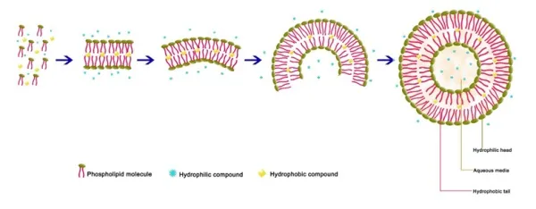
Figure 2. Formation of a unilamellar liposome and its basic structure.
What Are Liposomes Made Of?
The structural backbone of conventional liposomes is primarily composed of phospholipids, including glycerophospholipids and sphingophospholipids. Both contain hydrophilic head groups and hydrophobic tail regions.
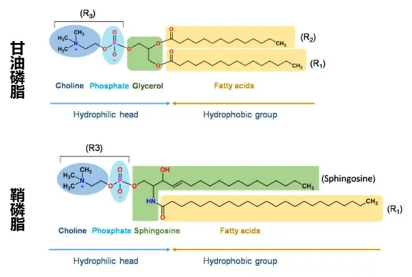
Figure 3. Schematic structures of glycerophospholipids and sphingophospholipids.
In an aqueous environment, phospholipids spontaneously arrange into bilayers through hydrophobic and other intermolecular interactions. The source also notes that cholesterol is commonly added to liposomal systems. Its roles can include promoting lipid-chain packing and bilayer formation, reducing membrane fluidity, limiting leakage of water-soluble compounds, reducing phospholipid loss, and improving structural stability.
How Are Liposomes Classified?
Liposomes can be classified in several ways, including by the number of lipid bilayers, particle size, surface charge, and surface modification.
1) Classification by Bilayer Number and Size
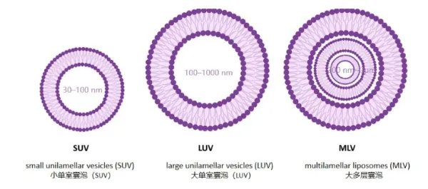
Figure 4. Schematic comparison of small unilamellar, large unilamellar and multilamellar vesicles.
Liposome Type | Bilayers | Typical Diameter |
Small unilamellar vesicles (SUVs) | 1 | 20-100 nm |
Large unilamellar vesicles (LUVs) | 1 | 100-400 nm |
Giant unilamellar vesicles (GUVs) | 1 | >=1 µm |
Multilamellar vesicles (MLVs) | >1 | 200 nm-3 µm |
2) Classification by Surface Modification
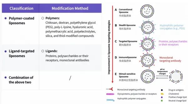
Liposome surfaces can also be modified with different components to alter properties such as stability, membrane interaction, or cellular permeability. The source illustrates conventional, stealth, targeted, immunoliposomal and stimulus-responsive concepts.
What Is Liposomal NMN?
Nicotinamide mononucleotide (NMN) can be incorporated into a liposomal system composed mainly of phospholipids and, depending on the formulation, cholesterol or other supporting components. The source describes Liposomal NMN as NMN enclosed within or associated with nanoscale lipid vesicles.
The formulation objective is to create a protective environment around NMN and potentially improve its stability and delivery characteristics. For a commercial ingredient, the actual NMN loading, particle characteristics, encapsulation or association efficiency, and storage stability should be evaluated for the specific material.
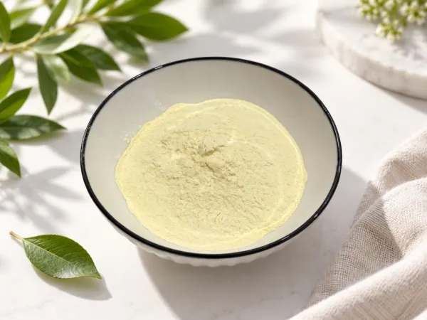
Figure 9. NMN powder shown in the source material.
2025 Comparative Human Study: Liposomal NMN vs. Non-Liposomal NMN
The source summarizes a double-blind controlled intervention study published in February 2025 in Annals of Clinical and Medical Case Reports. The study compared blood NAD+ responses after supplementation with Liposomal NMN, conventional non-liposomal NMN, or placebo.
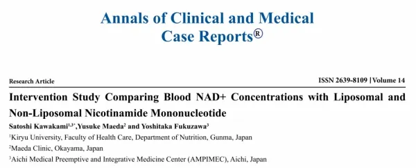
Figure 10. Article information for the 2025 study comparing Liposomal and Non-Liposomal NMN.
Study Design
The study enrolled 15 healthy men over 40 years of age. Participants were randomly divided into three groups and followed for a four-week daily intervention.
Group | Daily Intake | Intervention |
A - Placebo | 4 g maltodextrin, no NMN | 4 weeks |
B - Liposomal NMN | 350 mg/day | 4 weeks |
C - Non-Liposomal NMN | 350 mg/day conventional free NMN | 4 weeks |
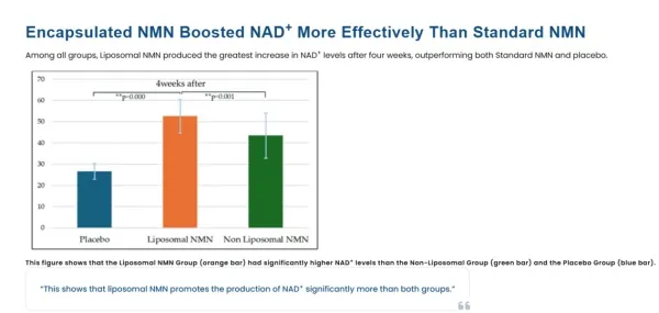
Figure 11. Summary chart in the source comparing the NAD+ response among placebo, Liposomal NMN and Non-Liposomal NMN.
Changes in NAD+ Over Time
The source includes individual group curves showing NAD+ measurements before intake, one hour after intake, after four weeks of supplementation, and at the follow-up point four weeks after supplementation had ended.
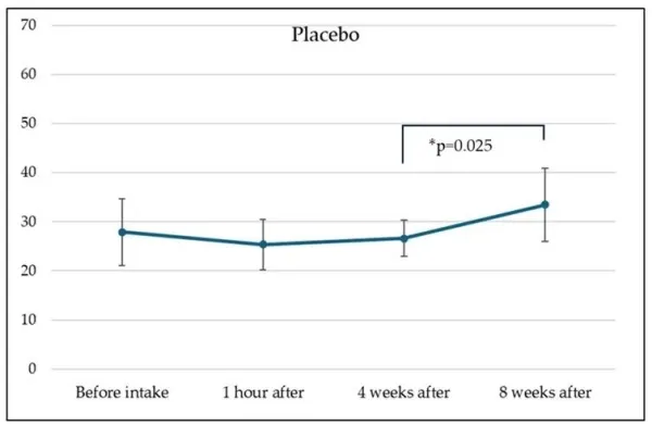
Figure 12. NAD+ levels over time in the placebo group.
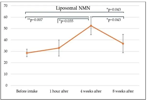
Figure 13. NAD+ levels over time in the Liposomal NMN group.
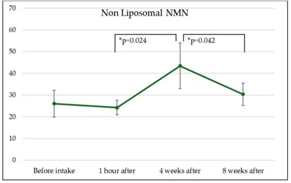
Figure 14. NAD+ levels over time in the Non-Liposomal NMN group.
Key Reported Result: 83.6% Increase in the Liposomal NMN Group
After four weeks of supplementation, the source reports that mean blood-cell NAD+ concentration in the Liposomal NMN group increased from 28.6 µM at baseline to 52.5 µM, representing an 83.6% increase.
The summary section of the source also states that the magnitude of the NAD+ increase in the Liposomal NMN group was 38% higher than that observed with conventional NMN.
Between-Group Statistical Comparisons at Week 4
Comparison | Reported p-value |
Liposomal NMN vs. Placebo | p = 0.000 |
Liposomal NMN vs. Non-Liposomal NMN | p = 0.001 |
Non-Liposomal NMN vs. Placebo | p = 0.545 |
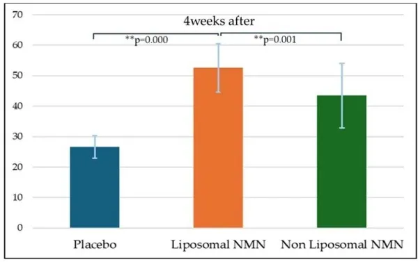
Figure 15. Between-group comparison of NAD+ after four weeks of intake.
In the source, p < 0.05 is treated as the threshold for statistical significance. Accordingly, the reported week-4 difference between Liposomal NMN and Non-Liposomal NMN was statistically significant (p = 0.001), whereas the reported difference between Non-Liposomal NMN and placebo was not statistically significant (p = 0.545).
What Happened After Supplementation Stopped?
All participants were assessed again four weeks after supplementation ended. The source reports that NAD+ concentration in the Liposomal NMN group remained significantly above its pre-intake baseline at this follow-up point (p = 0.043).
The source interprets this finding as evidence of a more persistent response. For a technical website article, the more precise conclusion is that the tested Liposomal NMN formulation showed a statistically significant elevation from baseline at the four-week post-intervention follow-up.
How Does the Source Explain the Difference?
The source attributes the stronger NAD+ response to the protective effect of the liposomal system, proposing that encapsulation may reduce NMN loss in the gastrointestinal tract and improve delivery.
It is important to distinguish this proposed mechanism from what the study directly measured. The reported clinical endpoint was blood NAD+ concentration; the source does not present direct measurements of intestinal NMN degradation, absolute absorption, cellular uptake, or pharmacokinetic bioavailability. Therefore, the clinical data are best described as evidence of a different NAD+ response with the tested formulation, while the precise mechanism should be supported by formulation-specific studies.
What Should Brands Evaluate in a Liposomal NMN Ingredient?
Actual NMN content per gram of the liposomal ingredient.
Phospholipid source and phospholipid concentration.
Complete carrier and excipient composition.
Particle-size distribution and the analytical method used.
PDI and zeta potential where applicable to the system.
Encapsulation or association efficiency, including how the value is defined and measured.
Stability of NMN and the liposomal system during storage.
Batch-specific COA, identity, purity and contaminant testing.
Whether clinical or technical evidence relates to the same or a sufficiently comparable liposomal formulation.
Liposomal NMN for Supplement Formulation
For finished-product development, Liposomal NMN may be used as a standalone ingredient or incorporated into broader healthy-aging formulas. A critical formulation point is the difference between the weight of the liposomal raw material and the amount of active NMN it delivers.
Liposomal powders contain phospholipids and may contain other carriers in addition to NMN. Therefore, capsule fill weight, bulk density and active-equivalent NMN dosage should be calculated before finalizing the formula. This becomes especially important in multi-ingredient capsule products where total fill capacity is limited.
Frequently Asked Questions
What is Liposomal NMN?
Liposomal NMN is NMN incorporated into a phospholipid-based vesicular delivery system rather than supplied only as conventional free NMN powder.
How is Liposomal NMN different from regular NMN?
The principal difference is the delivery format. Conventional NMN is supplied as free NMN, while Liposomal NMN combines NMN with a phospholipid-based system intended to protect and modify delivery behavior.
What did the 2025 study report?
The source reports that 15 healthy men over 40 were divided into placebo, Liposomal NMN and Non-Liposomal NMN groups. The NMN groups received 350 mg/day for four weeks.
How much did NAD+ increase in the Liposomal NMN group?
Mean NAD+ was reported to increase from 28.6 µM at baseline to 52.5 µM after four weeks, corresponding to an 83.6% increase.
Was Liposomal NMN significantly different from Non-Liposomal NMN?
At the four-week measurement, the source reports p = 0.001 for the Liposomal NMN versus Non-Liposomal NMN comparison.
Did the effect remain after supplementation stopped?
The source reports that NAD+ in the Liposomal NMN group remained significantly above baseline four weeks after supplementation ended (p = 0.043).
Does this prove that every Liposomal NMN product is more bioavailable?
No. The data relate to the specific formulation tested. Liposome composition, NMN loading, particle properties and manufacturing can differ substantially between products.
Conclusion
Liposomal NMN combines NMN with a phospholipid-based delivery platform. The source material provides a detailed overview of liposome structure and composition and highlights a 2025 human intervention study in which the tested Liposomal NMN formulation produced a stronger reported NAD+ response than the tested Non-Liposomal NMN formulation.
For brands and formulators, the most important question is not simply whether an ingredient is described as 'liposomal.' A credible Liposomal NMN ingredient should be supported by clear information on NMN loading, phospholipid composition, particle characteristics, encapsulation or association, stability and batch-level quality.
OriginBio
A reliable one stop nutrition supplement contract manufacturing partner
 EN
EN
 fr
fr  de
de  ru
ru 








 Call us on:
  Email Us:
  Office Add: